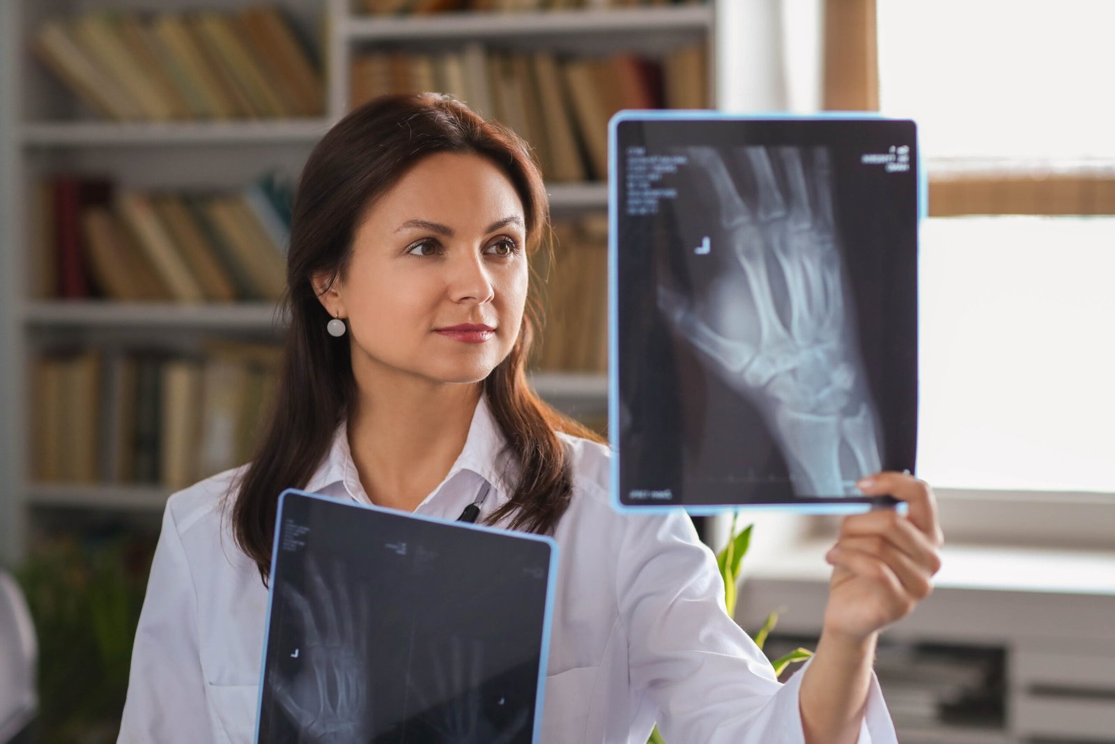
The X-Ray department at Vihaan Hospital and Research Centre is a critical diagnostic resource, equipped with advanced radiographic technology to aid in the accurate diagnosis of various medical conditions. Specializing in both routine and specialized X-ray imaging, the department caters to a wide range of diagnostic needs, from bone fractures and chest conditions to more complex studies like barium examinations and fluoroscopy-guided procedures. Staffed by experienced radiologists and skilled radiologic technologists, the department ensures high-quality imaging with a focus on patient safety and comfort. Utilizing digital radiography, the department offers rapid image processing, enhanced image quality, and reduced radiation exposure, contributing to efficient and effective patient care.
X – Ray F&Q's
An X-ray is a type of electromagnetic radiation used to create images of the inside of the body. It works by passing X-ray beams through the body, with different tissues absorbing or transmitting the rays differently. This creates an image on a detector, which shows the internal structures like bones, organs, and tissues.
X-rays are commonly used in medical diagnostics to identify fractures, dislocations, and bone deformities. They are also used to detect abnormalities in organs such as the lungs, heart, and abdomen, aiding in the diagnosis of conditions like pneumonia, heart disease, and gastrointestinal issues. Additionally, X-rays are used in dental imaging for detecting tooth decay and jaw problems.
X-rays differ from other imaging techniques like CT scans and MRIs in terms of the type of radiation used and the images produced. X-rays use ionizing radiation and produce 2D images primarily focused on bones and dense tissues. CT scans also use X-rays but produce detailed 3D images of soft tissues and organs. MRIs use magnetic fields and radio waves to produce highly detailed images of soft tissues and do not involve ionizing radiation.
Several precautions are taken to minimize radiation exposure during X-ray procedures. These include using lead aprons and thyroid shields to protect the patient’s body from unnecessary exposure, ensuring the X-ray machine is properly calibrated and operated by trained professionals, and using the lowest possible dose of radiation needed to obtain diagnostic images. Pregnant women are particularly cautious, and shielding of the abdomen is often used to protect the fetus.
Digital technology has improved X-ray imaging by providing faster image acquisition, higher image quality, and the ability to manipulate and store images digitally. Digital X-ray systems produce images that can be viewed immediately on computer screens, allowing for quick interpretation and reducing the need for repeat exposures. Additionally, digital images can be easily shared between healthcare providers and stored electronically for future reference.
Some limitations and risks associated with X-ray imaging include exposure to ionizing radiation, which carries a small risk of radiation-induced cancer, especially with repeated exposures. Additionally, X-rays may not provide detailed images of soft tissues, leading to limitations in diagnosing certain conditions. Pregnant women are particularly cautious about X-ray exposure due to potential risks to the fetus. Finally, some patients may be allergic to contrast agents used in certain types of X-ray procedures.


