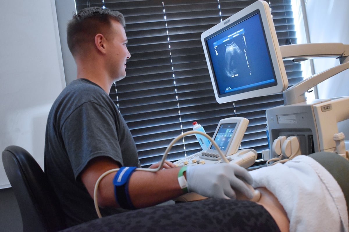
The Sonography department at Vihaan Hospital and Research Centre is a sophisticated facility offering a wide range of ultrasound imaging services. Equipped with the latest ultrasound technology, the department provides high-resolution images for accurate diagnosis and patient care. The experienced sonographers and radiologists specialize in abdominal, obstetric, pelvic, vascular, and musculoskeletal sonography, ensuring comprehensive diagnostic coverage. Emphasizing patient comfort and safety, the facility offers a serene and supportive environment. The department is integral in prenatal care, guiding interventional procedures, and aiding in the diagnosis of various medical conditions, making it an essential aspect of Vihaan Hospital’s commitment to advanced and compassionate healthcare.
Sonography F&Q's
Sonography, also known as ultrasound imaging, is a diagnostic medical procedure that uses high-frequency sound waves to produce images of the inside of the body. The sound waves are sent into the body using a device called a transducer. These waves bounce off tissues, organs, and fluids at different rates, and the echoes are captured by the transducer. A computer then uses these echoes to create an image that is displayed on a monitor.
Sonography is used in various medical fields for different purposes. It is commonly used for examining the fetus during pregnancy to monitor its development and health. It’s also used to diagnose conditions related to the abdomen, heart, blood vessels, kidneys, liver, gallbladder, pancreas, thyroid, and reproductive organs. Additionally, sonography can help guide needle biopsies and assess damage after an injury.
Sonography has several advantages, including being non-invasive and free from ionizing radiation, making it safer than X-rays and CT scans, especially for pregnant women and the fetus. It is also relatively low cost, widely available, and provides real-time imaging, which is beneficial for guiding procedures such as needle biopsies or when assessing the movement of internal organs.
Preparation for a sonography exam depends on the area being examined. For some abdominal scans, you may be asked to fast for several hours before the test or to drink water to fill your bladder for better visualization. For other types of ultrasound exams, no special preparation is needed. Your healthcare provider will give you specific instructions based on the type of ultrasound you are having.
Sonography can help in detecting and evaluating lumps or abnormalities in various organs and tissues in the body, which may indicate the presence of cancer. However, while sonography can be an important tool in the detection process, it cannot definitively diagnose cancer. Further tests, such as biopsies, are usually required to confirm a cancer diagnosis.
While sonography is a valuable diagnostic tool, it has limitations. It may not effectively image structures inside dense bone or parts of the body that hold air or gas, such as the lungs. In some cases, obesity can also hinder the quality of ultrasound images because the sound waves have difficulty penetrating deeper tissues. Additionally, sonography may not always distinguish between benign and malignant tumors, necessitating further diagnostic procedures for a definitive diagnosis.


